Effect of heat treatment duration on microstructure evolution and electrochemical degradation behavior of electrodeposited Fe-4.8Zn alloy
-
摘要: 以脉冲电沉积制备的 Fe-4.8Zn 合金为研究对象,在 450 ℃下进行不同保温时间热处理,系统研究了热处理时间对其显微组织、物相组成、元素分布、显微硬度及电化学降解行为的影响。结果表明,热处理促进了合金表面及截面组织的回复、重排与均匀化;XRD 结果显示,各样品均以 α-Fe 固溶体为主,热处理后衍射峰向高角度方向轻微偏移。合金中Fe与Zn分布均匀,未发生明显的元素再分布。随着保温时间延长,合金显微硬度逐渐升高,腐蚀电位逐步正移,腐蚀电流密度和腐蚀速率持续降低,电荷转移电阻逐渐增大。表明热处理时间对 Fe-4.8Zn 合金的组织结构、力学性能及电化学降解行为具有显著影响,可为 Fe-Zn 可降解血管支架材料的热处理调控提供试验依据。Abstract: Fe-4.8Zn alloy prepared by pulse electrodeposition was heat-treated at 450 ℃ for different holding times. The effects of heat treatment duration on the microstructure, phase composition, elemental distribution, microhardness, and electrochemical degradation behavior of the alloy were systematically studied. The results showed that heat treatment improved the recovery, rearrangement and homogenization of the microstructure on the surface and cross-section of the alloy. XRD analysis indicated that all samples were mainly composed of the α-Fe solid solution, and the diffraction peaks shifted slightly toward higher angles after heat treatment. The distributions of Fe and Zn were uniform, and no obvious elemental redistribution was observed. With increasing holding time, the microhardness of the alloy gradually increased, the corrosion potential shifted positively, the corrosion current density and corrosion rate decreased continuously, and the charge transfer resistance increased progressively. These results indicate that heat treatment duration has a significant effect on the microstructure, mechanical properties, and electrochemical degradation behavior of Fe-4.8Zn alloy, which provides an experimental support for the heat-treatment optimization of Fe-Zn alloys for biodegradable vascular stent applications.
-
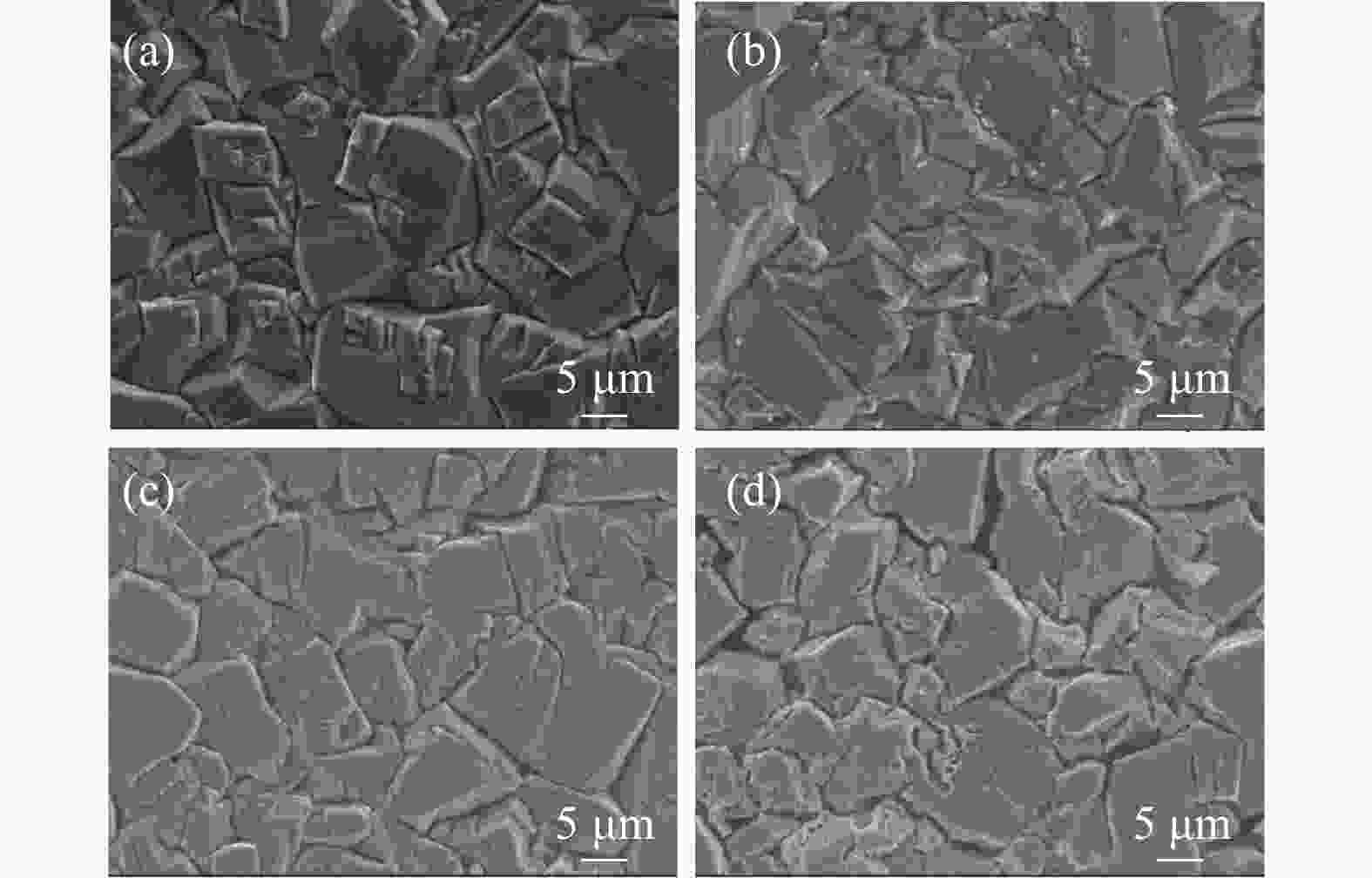
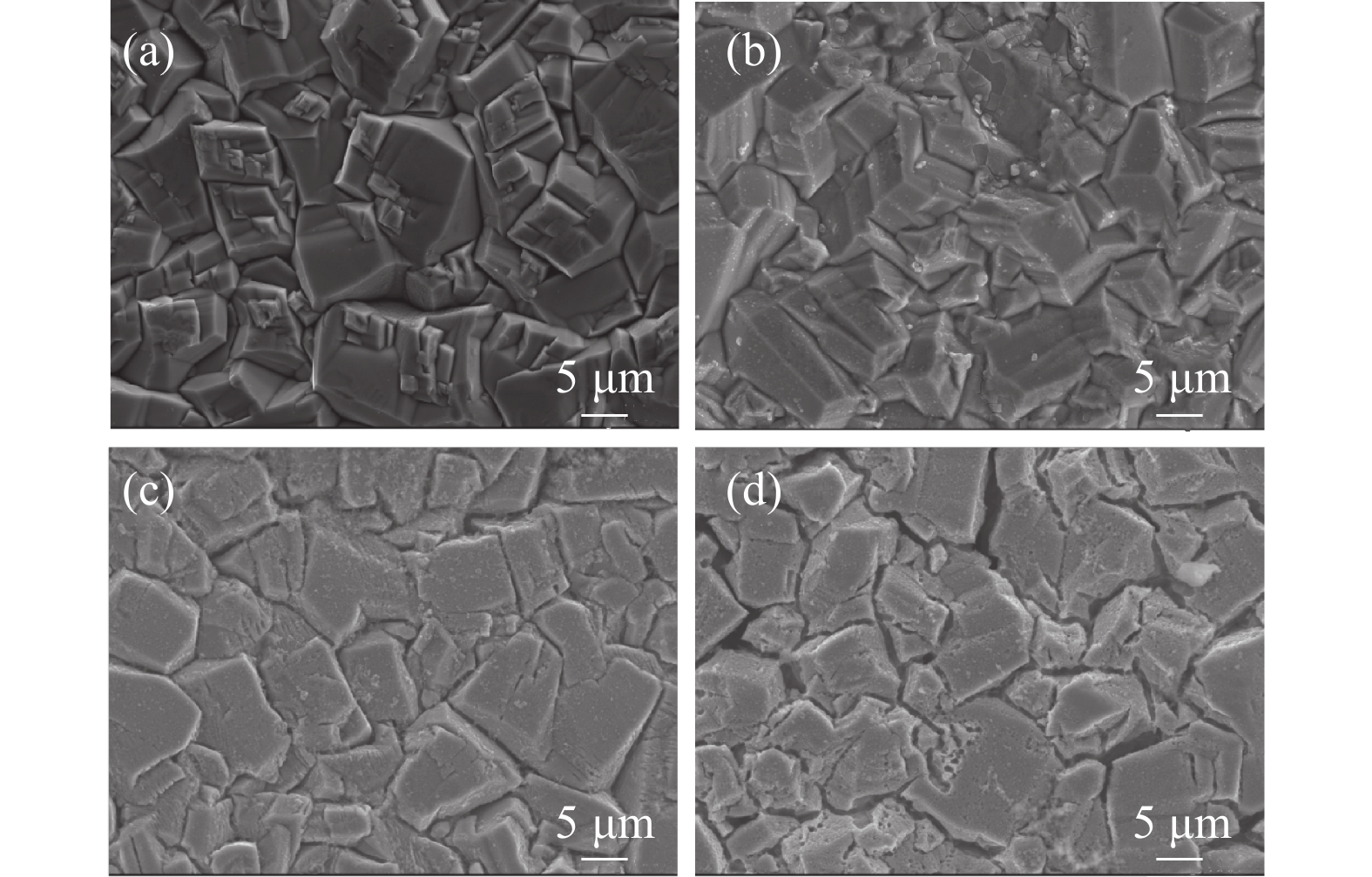
图 2 不同保温时间下Fe-4.8Zn的表面形貌
Figure 2. Surface morphology of Fe-4.8Zn under various holding times
(a) Untreated; (b) 20 min;(c) 40 min;(d) 60 min;
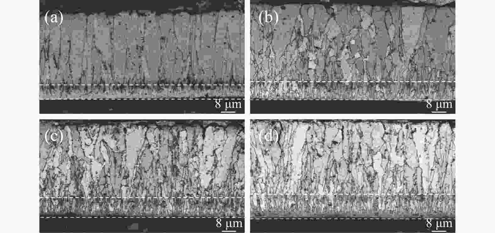
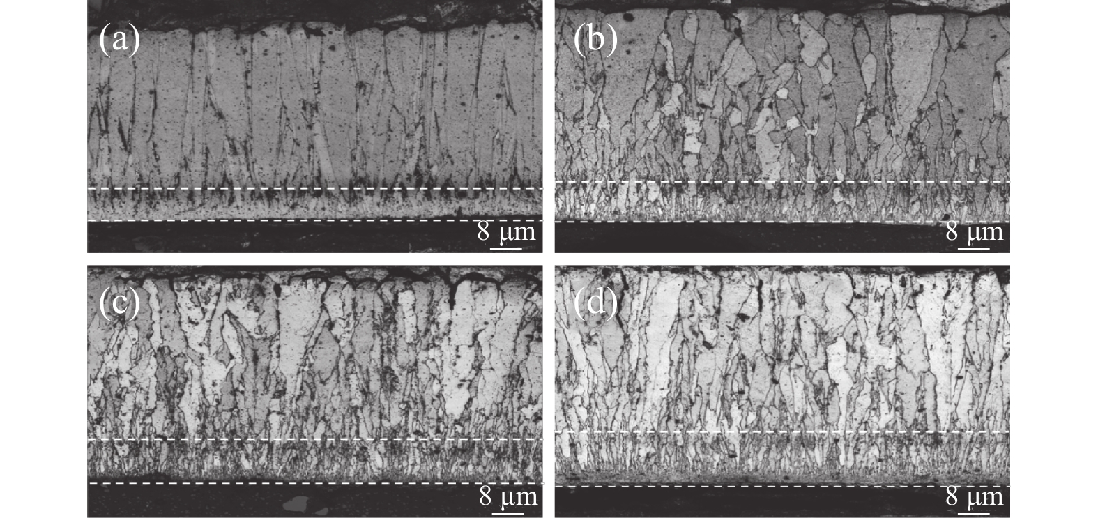
图 3 不同保温时间下Fe-4.8Zn的截面形貌
Figure 3. Cross-sectional morphology of Fe-4.8Zn alloy under various holding times
(b) Untreated; (b) 20 min;(c) 40 min;(d) 60 min;
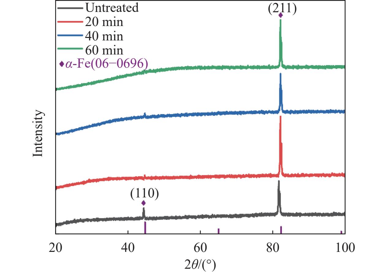
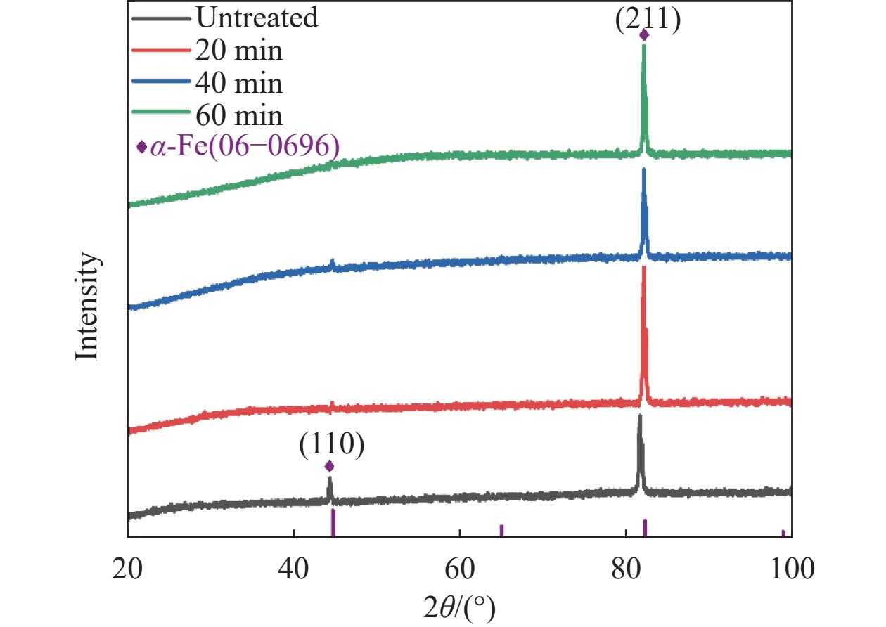
图 4 不同保温时间下Fe-4.8Zn的XRD图谱
Figure 4. XRD patterns of Fe-4.8Zn under various holding times
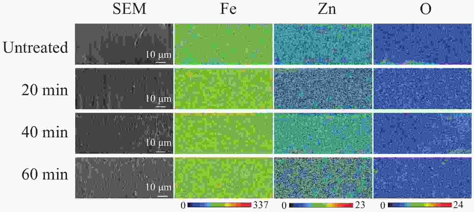
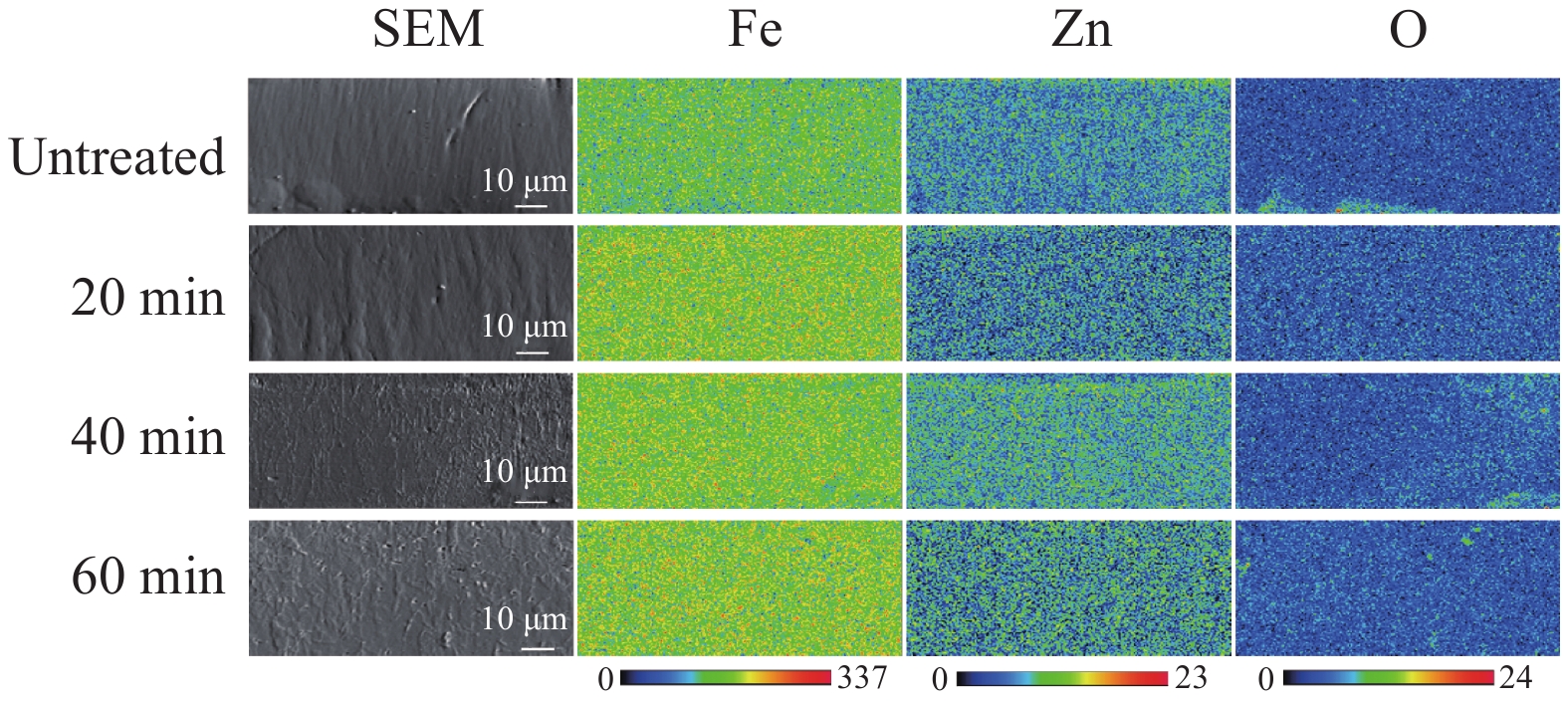
图 5 不同保温时间下Fe-4.8Zn的截面元素分布
Figure 5. Cross-sectional elemental distribution of Fe-4.8Zn under different holding times
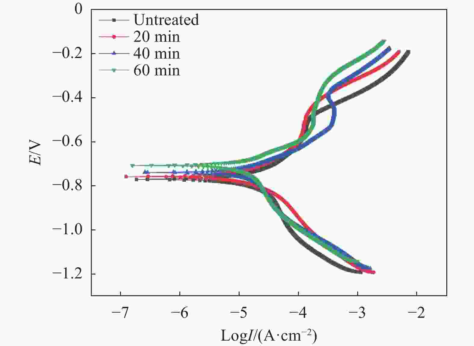
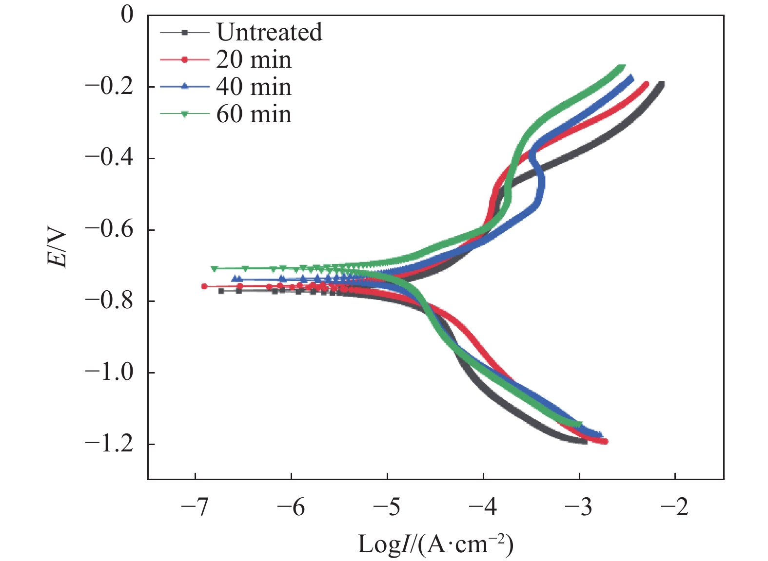
图 7 不同保温时间下Fe-4.8Zn在SBF溶液中的极化曲线
Figure 7. Polarization curves of Fe-4.8Zn in SBF solution under various holding times
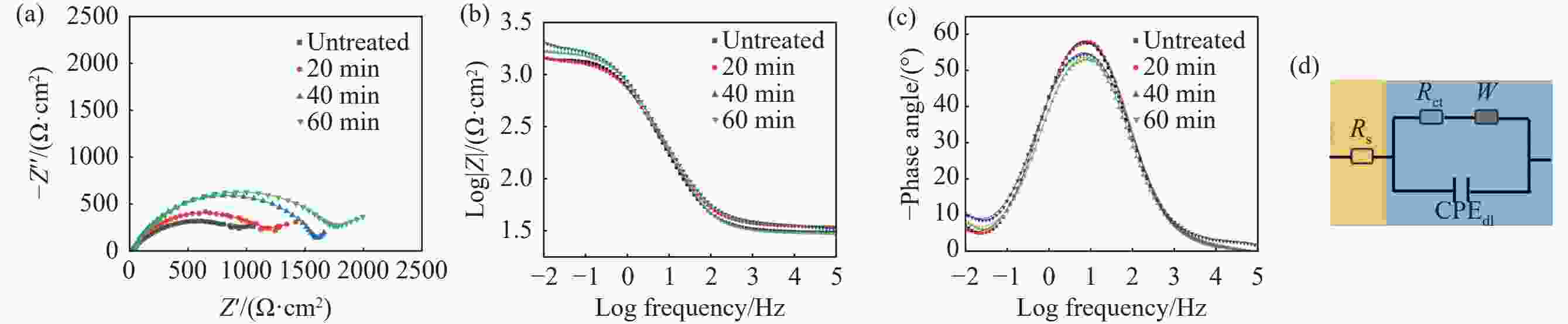
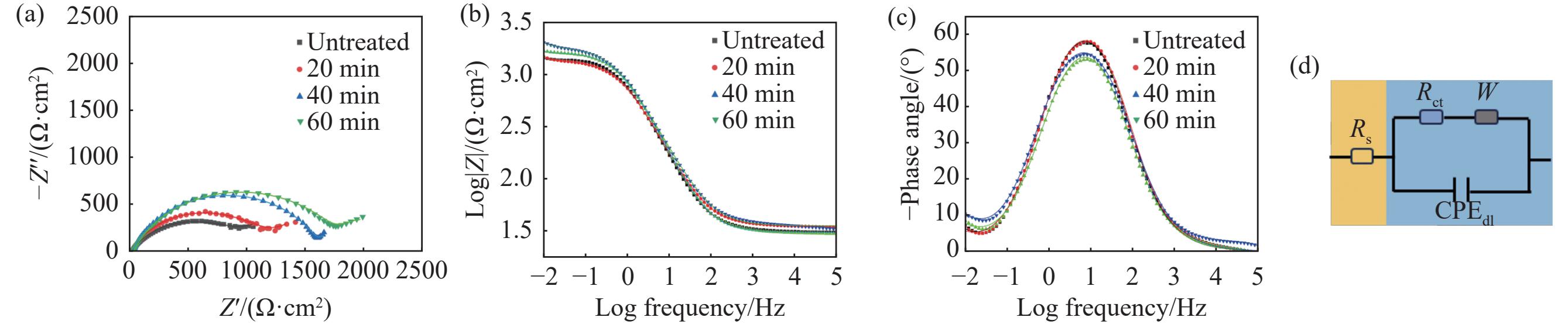
图 8 不同保温时间下Fe-4.8Zn在SBF溶液中的EIS结果
(a)Nyquist图谱;(b)(c)Bode图;(d)等效电路模型
Figure 8. EIS results of Fe-4.8Zn alloy in SBF solution under different holding times
表 1 电沉积Fe-Zn合金溶液组成
Table 1. Bath compositions of electrodeposition for Fe-Zn alloys
Chemicals Concentration/(g·L-1) Purity/% FeCl2·4H2O 253.00 99.0 ZnCl2 30.50 98.0 NaCl 45.00 AR MnCl2·4H2O 19.50 99.0 C6H8O6 5.00 AR C6H5Na3O7·2H2O 0.88 99.0 C6H4SO2NNaCO·2H2O 1.90 AR CH3(CH2)11OSO3Na 0.10 AR H3BO3 30.00 99.5 C3H4O4 5.00 98.0  下载: 导出CSV
表 2 电沉积Fe-4.8Zn合金的工艺参数
Table 2. Process parameters for the electrodeposition of Fe-4.8Zn alloy
Parameters ton+/ms toff+/ms ton−/ms toff−/ms Ip+/(A·dm-2) Ip−/(A·dm-2) N+ N− F/Hz setpoint 0.2 1.8 0.2 1.8 10 8 20 5 500
下载: 导出CSV
表 3 配制
1000 mL SBF所需的试剂及用量Table 3. Reagents and dosages for preparing 1000 mL SBF
Reagents Amount/g Purity/% NaCl 8.036 AR NaHCO3 0.352 AR KCl 0.225 AR K2HPO4·3H2O 0.230 AR MgCl2·6H2O 0.311 AR HCl* 40* CaCl2 0.293 AR Na2SO4 0.072 AR TRIS 6.063 99.8 注:*指浓度为1.0 mol/mL的HCl溶液,用量为40 mL。
下载: 导出CSV
表 4 从动电位极化曲线获得的电化学参数
Table 4. Electrochemical parameters obtained from potentiodynamic polarization curves
Times/min Ecorr /V Icorr /(μA·cm-2) CR/(mm·a-1) Untreated −0.769 17.431 0.205 20 −0.757 14.699 0.173 40 −0.737 13.310 0.157 60 −0.706 11.804 0.139
下载: 导出CSV
表 5 电化学阻抗谱拟合结果
Table 5. Fitting results of EIS
Times/min Rs/(Ω·cm−2) WR/(Ω·cm2) CPEdl×104/(F·cm2·s1-n) ndl Rct/(Ω·cm-2) Untreated 24.31 1257 6.99 0.72 922 20 29.14 1246 3.66 0.77 1128 40 30.51 1295 2.04 0.81 1578 60 35.40 1326 2.18 0.77 1749
下载: 导出CSV
-
[1] Zheng Jianfeng, Zheng Jilin, Lou Xiaoying. TCTAP A-017 long-term outcomes of percutaneous coronary intervention versus coronary artery bypass surgery in elderly patients with multivessel and/or left main coronary artery disease and diabetes mellitus: a systematic review and meta-analysis[J]. Journal of the American College of Cardiology, 2021, 77(14): S12-S13. [2] Byrne R A, Serruys P W, Baumbach A, et al. Report of a European Society of Cardiology-European Association of Percutaneous. Cardiovascular Interventions task force on the evaluation of coronary stents in Europe: executive summary[R]. European Heart Journal, 2015, 36(38): 2608-2620. [3] Gruntzig A. Transluminal dilatation of coronary-artery stenosis[J]. Lancet, 1978, 1(8058): 263. doi: 10.1007/978-3-642-46381-5_9 [4] Fischman D L, Leon M B, Baim D S, et al. A randomized comparison of coronary-stent placement and balloon angioplasty in the treatment of coronary artery disease. Stent restenosis study investigators[J]. The New England Journal of Medicine, 1994, 331(8): 496-501. doi: 10.1056/NEJM199408253310802 [5] Stone G W, Ellis S G, Cox D A, et al. A polymer-based, paclitaxel-eluting stent in patients with coronary artery disease[J]. The New England Journal of Medicine, 2004, 350(3): 221-231. [6] Shen Y H, Yu X, Cui J, et al. Development of biodegradable polymeric stents for the treatment of cardiovascular diseases[J]. Biomolecules, 2022, 12(9): 1245. [7] Lee H I, Heo Y, Baek S W, et al. Multifunctional biodegradable vascular PLLA scaffold with improved X-ray opacity, anti-inflammation, and re-endothelization[J]. Polymers, 2021, 13(12): 1979. [8] Wang Peijiang, Ferralis N, Conway C, et al. Strain-induced accelerated asymmetric spatial degradation of polymeric vascular scaffolds[J]. Proceedings of the National Academy of Sciences of the United States of America, 2018, 115(11): 2640-2645. [9] Bowen P K, Shearier E R, Zhao Shan, et al. Biodegradable metals for cardiovascular stents: from clinical concerns to recent Zn-alloys[J]. Advanced Healthcare Materials, 2016, 5(10): 1121-1140. [10] Haude M, Ince H, Abizaid A, et al. Safety and performance of the second-generation drug-eluting absorbable metal scaffold in patients with de-novo coronary artery lesions (BIOSOLVE-II): 6 month results of a prospective, multicentre, non-randomised, first-in-man trial[J]. Lancet, 2016, 387(10013): 31-39. [11] Heublein B, Rohde R, Kaese V, et al. Biocorrosion of magnesium alloys: a new principle in cardiovascular implant technology?[J]. Heart, 2003, 89(6): 651-656. [12] Liu Dexiao, Yang Ke, Chen Shanshan. Development and future trends of protective strategies for magnesium alloy vascular stents[J]. Materials, 2023, 17(1): 68. [13] Mostaed E, Sikora-Jasinska M, Drelich J W, et al. Zinc-based alloys for degradable vascular stent applications[J]. Acta Biomaterialia, 2018, 71: 1-23. [14] Mostaed E, Sikora-Jasinska M, Ardakani M S, et al. Towards revealing key factors in mechanical instability of bioabsorbable Zn-based alloys for intended vascular stenting[J]. Acta Biomaterialia, 2020, 105: 319-335. [15] Gasior G, Szczepański J, Radtke A. Biodegradable iron-based materials—What was done and what more can be done?[J]. Materials, 2021, 14(12): 3381. [16] Obayi C S, Tolouei R, Mostavan A, et al. Effect of grain sizes on mechanical properties and biodegradation behavior of pure iron for cardiovascular stent application[J]. Biomatter, 2016, 6(1): e959874. [17] Zhang Yang, Roux C, Rouchaud A, et al. Recent advances in Fe-based bioresorbable stents: Materials design and biosafety[J]. Bioactive Materials, 2024, 31: 333-354. [18] Hermawan H, Dubé D, Mantovani D. Degradable metallic biomaterials: design and development of Fe-Mn alloys for stents[J]. Journal of Biomedical Materials Research Part A, 2010, 93(1): 1-11. [19] Feng Qimao, Zhang Deyuan, Xin Chaohua, et al. Characterization and in vivo evaluation of a bio-corrodible nitrided iron stent[J]. Journal of Materials Science: Materials in Medicine, 2013, 24(3): 713-724. [20] O'neal S L, Zheng Wei. Manganese toxicity upon overexposure: a decade in review[J]. Current Environmental Health Reports, 2015, 2(3): 315-328. [21] Hermawan H. Updates on the research and development of absorbable metals for biomedical applications[J]. Progress in Biomaterials, 2018, 7(2): 93-110. [22] Yang Hongtao, Jia Bo, Zhang Zechuan, et al. Alloying design of biodegradable zinc as promising bone implants for load-bearing applications[J]. Nature Communications, 2020, 11(1): 401. [23] Huang Tao, Zheng Yufeng, Han Yong. Accelerating degradation rate of pure iron by zinc ion implantation[J]. Regenerative Biomaterials, 2016, 3(4): 205-215. [24] Okamoto H. Fe-Zn (Iron-Zinc)[J]. Journal of Phase Equilibria and Diffusion, 2007, 28(3): 317-318. [25] Xu Yanan, Wang Weiqiang, Yu Fengyun, et al. Effects of pulse frequency and current density on microstructure and properties of biodegradable Fe-Zn alloy[J]. Journal of Materials Research and Technology, 2022, 18: 44-58. [26] Yang S K, Yu F Y, Wang W Q, et al. Biodegradable Fe-Zn alloy microtubes with good mechanical properties, degradability, and cytocompatibility prepared by electrodeposition[J]. Advanced Functional Materials, 2025, 35(28): e04797. [27] Xu Yanan, Wang Weiqiang, Yu Fengyun, et al. The enhancement of mechanical properties and uniform degradation of electrodeposited Fe-Zn alloys by multilayered design for biodegradable stent applications[J]. Acta Biomaterialia, 2023, 161: 309-323. [28] Wang Y C, Zhao D P, Li S K, et al. The heat treatment effect on microstructure and dynamic mechanical properties of electroformed nanocrystalline Ni–W alloy[J]. Materials Science and Engineering: A, 2012, 547: 104-109. [29] Moravej M, Prima F, Fiset M, et al. Electroformed iron as new biomaterial for degradable stents: Development process and structure–properties relationship[J]. Acta Biomaterialia, 2010, 6(4): 1726-1735. [30] Wang Qing, Li Zhen, Pang Shujie, et al. Coherent precipitation and strengthening in compositionally complex alloys: A review[J]. Entropy, 2018, 20(11): 878. [31] Hillel G, Meshi L, Kalabukhov S, et al. Shock wave characterization of precipitate strengthening of pH 13–8 Mo stainless steel[J]. Acta Materialia, 2020, 187: 176-185. -

计量
- 文章访问数: 0
- HTML全文浏览量: 0
- PDF下载量: 0
- 被引次数: 0